| 症例3 40歳代 男性 |
|
診断と解説
|
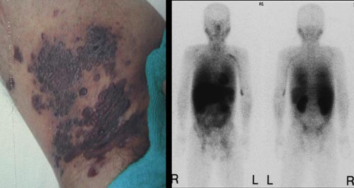
病理:kaposi's sarcoma |
診断 |
|
Kaposi's sarcoma(KS)
|
|
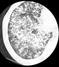
Kaposi's sarcoma of the lung
|
| ・画像所見のまとめ 鑑別診断へ |
| ・症例提示へ |
|
|
Moderator : 松尾 周也 |