|
第303回 東京レントゲンカンファレンス
2008年03月27日開催
|
| 症例1 70歳代 男性 |
右上肢の血圧が高い
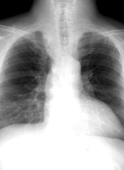
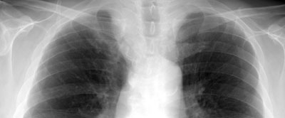
胸部単純写真所見
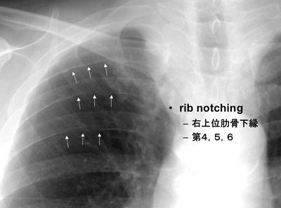
rib notching
右上位肋骨下縁
第4,5,6
上縦隔右縁の突出
肺尖部まで
気管右側壁を圧排
右腕頭動脈の拡張?
頸部右側の筋束の輪郭の不鮮明 |
|
Rib notching on inferior margin
Minimal scalloping to deep ridges along the neurovascular grooves
- Arterial; dilatation of intercostal artery as collateral to descending aorta or pulmonary artery
Aorta; Coactation, Atherothrombosis
Subclavian a.; Blalock-Taussig shunt
Pulmonary a.; PA stenosis or occlusion, Tetralogy of Fallot,
- Venous; dilatation of intercostal veins
AVM of chest wall, SVC obstruction
- Neurogenic;
Intercostal neuroma, NF, Poliomyelitis/quadri-/paraplegia
- Osseous;
hyperparathyroidism, Thalassemia, M-N Sx
Wolfgang Dahnert . Radiology Review Mannual
|
| |
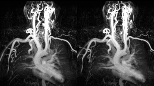
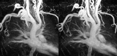
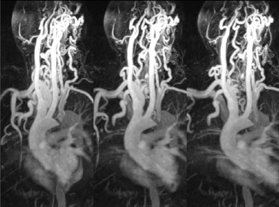
造影MRA
両側とも鎖骨下動脈から上腕動脈近位側には狭窄〜閉塞は認めない
|
|
|
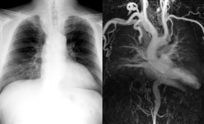
左鎖骨下動脈起始部レベルで大動脈縮窄
側副血流:右肋間動脈
右鎖骨下動脈→右上胸動脈、胸肩峰動脈、外側胸動脈→右肋間動脈→胸部下行大動脈 |
最終診断:
- 大動脈縮窄(症) Coarctation of the aorta
左鎖骨下動脈起始部閉塞(高度狭窄)
鎖骨下動脈盗血症候群
- Rib notching
Suggesting intercostal artery dilatation as collateral circulation into the thoracic aorta.
|
大動脈縮窄症 Coarctation of the aorta
単純型大動脈縮窄症
- Localized coarctation
- Adult type
- Juxtaductal type
HTx. in the upper ext.
Headache
Renovaascular HTx.
大動脈縮窄症複合
- Tubular hypoplasia
- Infantile type
- Diffuse type
Coexistent cardiac anomaly; PDA, VSD
CHF in neonate in 50%
Cyanosis in the lower ext.
病因
- 先天性
胎内感染・胎児期血行動異常が示唆
先天性風疹症候群の16%に合併
- 後天性
大動脈炎症候群
大動脈瘤
大動脈解離
動脈管からみた病態
- 管前型
下行大動脈への血流が右心系から動脈管を介して供給される。
- 管後型
下行大動脈への血流が左心系から側副血流供給される。
|
Rib Notching in Coactation
- Arterial; dilatation of intercostal artery as collateral to descending aorta or pulmonary artery
- 75%, mostly in adult type
- 3rd-8th ribs, especially in 3rd and 4th ribs
- Central to lateral thirds of posterior rib
|
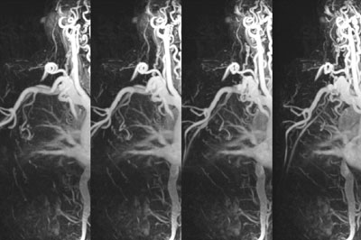
右肋間動脈の拡張
頸横動脈・肩甲横動脈・肩甲下動脈・下甲状腺動脈→肋間動脈→下行大動脈
|
|
胸部単純写真:造影MRAとの比較
上縦隔右縁の突出、とくに右鎖骨下縁よりも上部は、血流量増加にともなう右腕頭動脈から鎖骨下動脈近位側の拡張を反映している。 |
|
症例提示ページへ
|
|
|
|
|
|