|
第303回 東京レントゲンカンファレンス
2008年03月27日開催
|
| 症例8 20歳代 男性 |
画像所見のまとめ
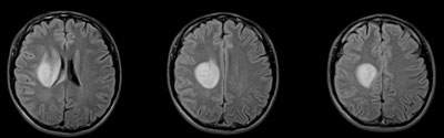
CT:右前頭頂葉白質から右内外包に広がるlow density lesionを認める。中心部分にenhancementを認める。
MRI:CT同様右前頭頂葉白質に脳梁と接する4cm程度の腫瘤を認める。また連続するように内外包の白質にそった病変の広がりを示し,側頭葉鈎部白質まで達する。
DWIで淡いhigh signalを示し,中心部に軽度の造影効果を認める。
Midline shiftはごく軽度で,Mass effectはあまり目立たない。
|
鑑別診断
腫瘍性病変:
Glioma(Oligodendroglioma, astrocytoma), Malignant lymphoma, Metastatic tumor
脱髄疾患:
Multiple sclerosis, ADEM, PML
感染症:
真菌感染など
脳血管障害:
Brain infarction(血栓塞栓,血管炎など)
|
|
入院時FLAIR |
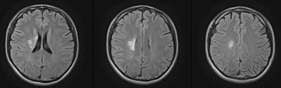
ステロイド投与1ヶ月後FLAIR
|
診断:Multiple Sclerosis
|
Large (2cm or more) tumefactive demyelinating plaques
- Well-demarcated, notably found in the white matter.
- CT: Hypodense
- MRI: T2WI high, T1WI low, rim-enhancement type is more common in the tumefactive variant than in the standard MS.
Neuroradiology 1996;38:560-565 Br J Radiol 2004;77:153-156
Helpful diagnostic feature of tumefactive MS
- Multiplicity of lesions.
- Typical periventricular location.
- Relative lack of mass effect or vasogenic edema.
- MR spectroscopy may be useful to differentiate from tumor (reduced NAA without corresponding elevation of choline peak relative to creatine peak).
|
|
症例提示ページへ
|
|
|
|
|
|