
2010年4月22日
線維形成性小円形細胞性腫瘍 |
画像所見
腹腔内に多発する腫瘤(脾門部、膀胱後部、ダグラス窩)
原発不明
ひとつひとつの腫瘤が比較的大きい
単純CTで低濃度な領域があり、同部位の造影効果は比較的弱い
リンパ節腫大
腫瘤内部に小さな石灰化
胸部に異常なし
臨床所見
若年
男性
腫瘍マーカー CA125
NSE 軽度上昇
アスベスト(-)
鑑別診断
Peritoneal Neoplasms
・Mesothelioma
・Omental / Peritoneal carcinomatosis (carcinoma of stomach, colon pancreas)
・Serous papillary carcinoma
・Desmoplastic small round cell tumor
・Malignant mesenchymal tumors
・Liposarcoma |
||
Others |
| 開腹生検 |
|
| 腫瘍は白色で充実性、弾性硬。割面では中心部に出血を伴った白色調の腫瘤。 |
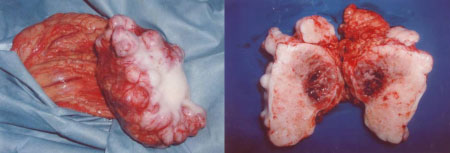
| 小型円形の腫瘍細胞が充実性胞巣状に増殖しており、発達した線維形成性の間質を伴って大小の島状に存在。 免疫組織染色検査ではDesmin,Vimentin,NSE,Cytokeratin,MIB-1 が陽性。 |
| Bellah R. Am J Roentgenol. 2005 ;184:1910-4 |
Small round cell tumor
・病理学的に構成細胞が小型で円形を示す腫瘍の総称。(small round cell tumorという腫瘍が存在するわけではない)
・確定診断のためには分子病理学的検索が必須。
・一般に細胞密度が高く、増殖能も高く、予後不良。
Small round cell tumorの範疇に入る腫瘍
・Ewing肉腫 |
・リンパ性白血病 |
Desmoplastic small round cell tumor(線維形成性小円形細胞性腫瘍)
・若年男性 臨床症状は腹痛,腹部膨満,腫瘤触知。
・確立した治療法なく予後不良(5年生存率35%)。
・血清腫瘍マーカーではCA125,NSE が軽度上昇することがある。
Desmoplastic small round cell tumorの画像所見
・大網や漿膜に接する円形、類円形の腫瘤(100%)
・10cm以上の腫瘤がある(91%)
・膀胱後部、直腸子宮窩に位置(82%)
・斑状・微小石灰化(55%)
・腹水(64%)
・肝転移(55%)
・リンパ節腫大(55%)
・腸閉塞(18%)
・単純CTで低濃度を示す領域があり、造影効果は弱く、内部に壊死部分を伴うことが多い(73%)
Bellah R. Am J Roentgenol. 2005 ;184:1910-4.
参考文献
・臨床画像 2010年 vol.26 No.1
・Tateishi U, et al. Desmoplastic small round cell tumor: imaging findings associated with clinicopathologic features. J Comput Assist Tomogr. 2002;26(4):579-83.
・Bellah R, et al. Desmoplastic small round cell tumor in the abdomen and pelvis: report of CT findings in 11 affected children and young adults. AJR. 2005;184(6):1910-4.
・Pickhardt PJ, et al.Primary neoplasms of peritoneal and sub-peritoneal origin: CT findings.Radiographics. 2005;25(4):983-95.
・Jeong YJ, et al. Neoplastic and nonneoplastic conditions of serosal membrane origin: CT findings. Radiographics. 2008;28(3):801-17
・Bahrami A, et al. Undifferentiated tumor: true identity by immunohistochemistry. Arch Pathol Lab Med. 2008;132(3):326-48.
| ≪≪症例提示へ戻る |