
| 肺サルコイドーシス pulmonary sarcoidosis |
| 鑑別診断 |
・膠原病合併のNSIPと肺気腫
・肺サルコイドーシスと肺気腫
経過
•経気管支肺生検を施行
| 画像所見 |
・気管支血管束や小葉間隔壁・胸膜などに結節
・中・上葉優位に分布
・肺の線維化所見(網状影, 牽引性気管支拡張, 嚢胞)
・リンパ節腫大Brauner MW, Grenier P, Mompoint D, Lenoir S, de Crémoux H. Pulmonary sarcoidosis: evaluation with high-resolution CT. Radiology 1989;172(2): 467–471. 29.
Müller NL, Kullnig P, Miller RR. The CT findings of pulmonary sarcoidosis: analysis of 25 patients. AJR Am J Roentgenol 1989;152(6):1179–1182.
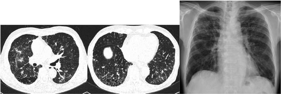 |
胸部単純写真では 上肺野優位であることが明瞭 |
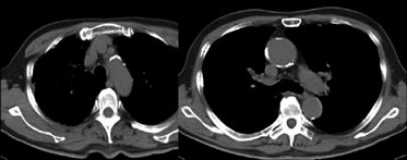 |
リンパ節は腫大していない |
Staging
| Stage | CXR Findings | 頻度 |
| 0 | No abnormalities | 5-10% |
| I | Lymphadenopathy | 50% |
| II | Lymphadenopathy + infiltration | 25-30% |
| III | Infiltration | 10-12% |
| IV | Fibrosis | 5% |
| Stageが上がるほど 肺機能が低下する傾向がある |
Siltzbach LE. Sarcoidosis: clinical features and management. Med Clin North Am 1967;51(2):483–502.
すりガラス影
・斑状に見られることが多い
・40%程度の症例で見られる
・結節の癒合や肺線維症によって生じる
Criando E. et al. Pulmonary Sarcoidosis: Typical and Atypical Manifestations at High-Resolution CT with Pathologic Correlation. Radiographics2010;30:1567-86
参考文献
- Brauner MW, Grenier P, Mompoint D, Lenoir S, de Cremoux H. Pulmonary sarcoidosis: evaluation with high-resolution CT.Radiology 1989;172(2): 467?471. 29.
- Muller NL, Kullnig P, Miller RR. The CT findings of pulmonary sarcoidosis: analysis of 25 patients. AJR Am J Roentgenol 1989;152(6):1179?1182.
|