
第 402 回 東京レントゲンカンファレンス[2023年6月22日]
| 症例4 30歳代 女性:過長月経 |
| 低悪性度子宮内膜間質肉腫 low-grade endometrial stromal sarcoma |
| 鑑別診断 |
筋層内嚢胞性腫瘤
・変性子宮筋腫
・子宮内膜間質腫瘍
・アデノマトイド腫瘍
・粘液癌転移
・Cystic adenomyosis(or ACUM)
筋層構造を保ち広がる境界不明瞭な腫瘤
・低悪性度子宮間質肉腫 ESS
・子宮腺筋症
脈管構造に沿う腫瘍の進展
・低悪性度子宮間質肉腫 ESS
・静脈内平滑筋腫症
1. Himoto Y et al. Sci Rep. 2021 Sep 27;11(1):19124.
| 子宮内膜間質肉腫 ESS |
・増殖期の子宮内膜間質細胞に似た間葉系腫瘍.
・子宮悪性腫瘍の約0.2%(子宮肉腫の10~15%).
・好発年齢40-55歳.
・晩期再発がしばしば報告される.
・High-grade:64歳>Low-grade:42歳(中央値).
・晩期再発を防ぐため、子宮全摘術に加え、両側卵巣摘出術が推奨される.
・術後にホルモン治療が行われる.
| WHO2020 Endometrial stromal tumours (ESTs) |
| Endometrial Stromal Nodules (ESNs) |
| Low-Grade Endometrial Stromal Sarcomas (LG-ESSs) |
| High-Grade Endometrial Stromal Sarcomas (HG-ESSs) |
| Undifferentiated Uterine Sarcomas (UUSs) |
2. D'Angelo E, Prat J. Gynecol Oncol. 2010 Jan;116(1):131-9.
3. Huang YL, et al. Cancer Imaging. 2019 Sep 12;19(1):63.
4. 子宮体がん治療ガイドライン2018年版. 金原出版: 195-198.
| 子宮内膜間質肉腫 ESS 画像所見 |
・筋層内に腫瘤形成 and/or 粘膜下にポリープ状。
・境界明瞭でT2WIで低信号rim or 筋層浸潤し境界不明瞭。
・腫瘍内に取り残された子宮筋層 →T2WIで帯状低信号やworm-likeな結節の広がり。
・靭帯や脈管に沿った進展。
・拡散強調像で高信号。
5. Koyama T, et al. AJR. 1999 Sep;173(3):767-72.
1. Himoto Y et al. Sci Rep. 2021 Sep 27;11(1):19124.・嚢胞はまれとされていたが、最近では少なくないと報告されている。
・Low-grade ESSと子宮筋腫を比較した研究で嚢胞はlow-grade ESSと関連があると示唆された。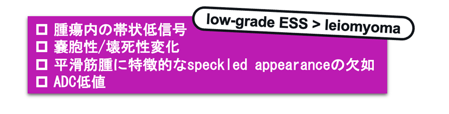
6. Park GE, et al. Ultrasonography. 2016 Apr;35(2):124-30.
7. Pérez-Montiel D, et al. Ann Diagn Pathol. 2004 Aug;8(4):213-8.
1. Himoto Y et al. Sci Rep. 2021 Sep 27;11(1):19124. ・FDG集積の報告は少ない。
・SUVmaxの値もばらばら(本例はSUVmax=2.62)。
・FDG高集積のESNの報告もあり。
8. Yamamoto M, et al. Oncotarget. 2017 Apr 4;8(14):22581-22589.
9. Umesaki N, et al. Gynecol Oncol. 2001 Mar;80(3):372-7.
10. Fujiishi K, et al. Diagn Pathol. 2019 Oct 15;14(1):110.
11. Maruyama S, et al. Case Rep Obstet Gynecol. 2015;2015:540283.
| Take home message |
・嚢胞を伴うlow-grade ESSは最近ではまれではないと考えられており、子宮内嚢胞性腫瘤の鑑別診断として挙げられる。
・病変の所在、進行状態で鑑別診断は変わってくる。
・特徴的な画像所見をとらえることで診断が可能と考えられる。
参考文献
- Himoto Y, Kido A, Sakata A,et al. Differentiation of uterine low-grade endometrial stromal sarcoma from rare leiomyoma variants by magnetic resonance imaging. Sci Rep. 2021 Sep 27;11(1):19124. doi: 10.1038/s41598-021-98473-z. PMID: 34580348; PMCID: PMC8476551.
- D'Angelo E, Prat J. Uterine sarcomas: a review. Gynecol Oncol. 2010 Jan;116(1):131-9. doi: 10.1016/j.ygyno.2009.09.023. Epub 2009 Oct 23. PMID: 19853898.
- Huang YL, Ueng SH, Chen K,et al. Utility of diffusion-weighted and contrast-enhanced magnetic resonance imaging in diagnosing and differentiating between high- and low-grade uterine endometrial stromal sarcoma. Cancer Imaging. 2019 Sep 12;19(1):63. doi: 10.1186/s40644-019-0247-z. PMID: 31514752; PMCID: PMC6739916.
- 子宮体がん治療ガイドライン2018年版. 金原出版: 195-198.
- Koyama T, Togashi K, Konishi I, et al. MR imaging of endometrial stromal sarcoma: correlation with pathologic findings. AJR Am J Roentgenol. 1999 Sep;173(3):767-72. doi: 10.2214/ajr.173.3.10470920. PMID: 10470920.
- Park GE, Rha SE, Oh SN,et al. Ultrasonographic findings of low-grade endometrial stromal sarcoma of the uterus with a focus on cystic degeneration. Ultrasonography. 2016 Apr;35(2):124-30. doi: 10.14366/usg.15045. Epub 2015 Oct 5. PMID: 26537303; PMCID: PMC4825207.
- Pérez-Montiel D, Salmeron AA, Domínguez Malagon H. Multicystic endometrial stromal sarcoma. Ann Diagn Pathol. 2004 Aug;8(4):213-8. doi: 10.1053/j.anndiagpath.2004.04.004. PMID: 15290672.
- Yamamoto M, Tsujikawa T, Yamada S, et al. 18F-FDG/18F-FES standardized uptake value ratio determined using PET predicts prognosis in uterine sarcoma. Oncotarget. 2017 Apr 4;8(14):22581-22589. doi: 10.18632/oncotarget.15127. PMID: 28186981; PMCID: PMC5410246.
- Umesaki N, Tanaka T, Miyama M,et al. Positron emission tomography with (18)F-fluorodeoxyglucose of uterine sarcoma: a comparison with magnetic resonance imaging and power Doppler imaging. Gynecol Oncol. 2001 Mar;80(3):372-7. doi: 10.1006/gyno.2000.6081. PMID: 11263934.
- Fujiishi K, Nagata S, Kano R,et al. JAZF1-SUZ12 endometrial stromal sarcoma forming subserosal masses with extraordinary uptake of fluorodeoxyglucose on positron emission tomography: a case report. Diagn Pathol. 2019 Oct 15;14(1):110. doi: 10.1186/s13000-019-0897-y. PMID: 31615558; PMCID: PMC6792225.
- Maruyama S, Sato Y, Satake Y, et al. Diffusion-Weighted MRI and FDG-PET in Diagnosis of Endometrial Stromal Nodule. Case Rep Obstet Gynecol. 2015;2015:540283. doi: 10.1155/2015/540283. Epub 2015 Jan 28. PMID: 25694838; PMCID: PMC4324744.